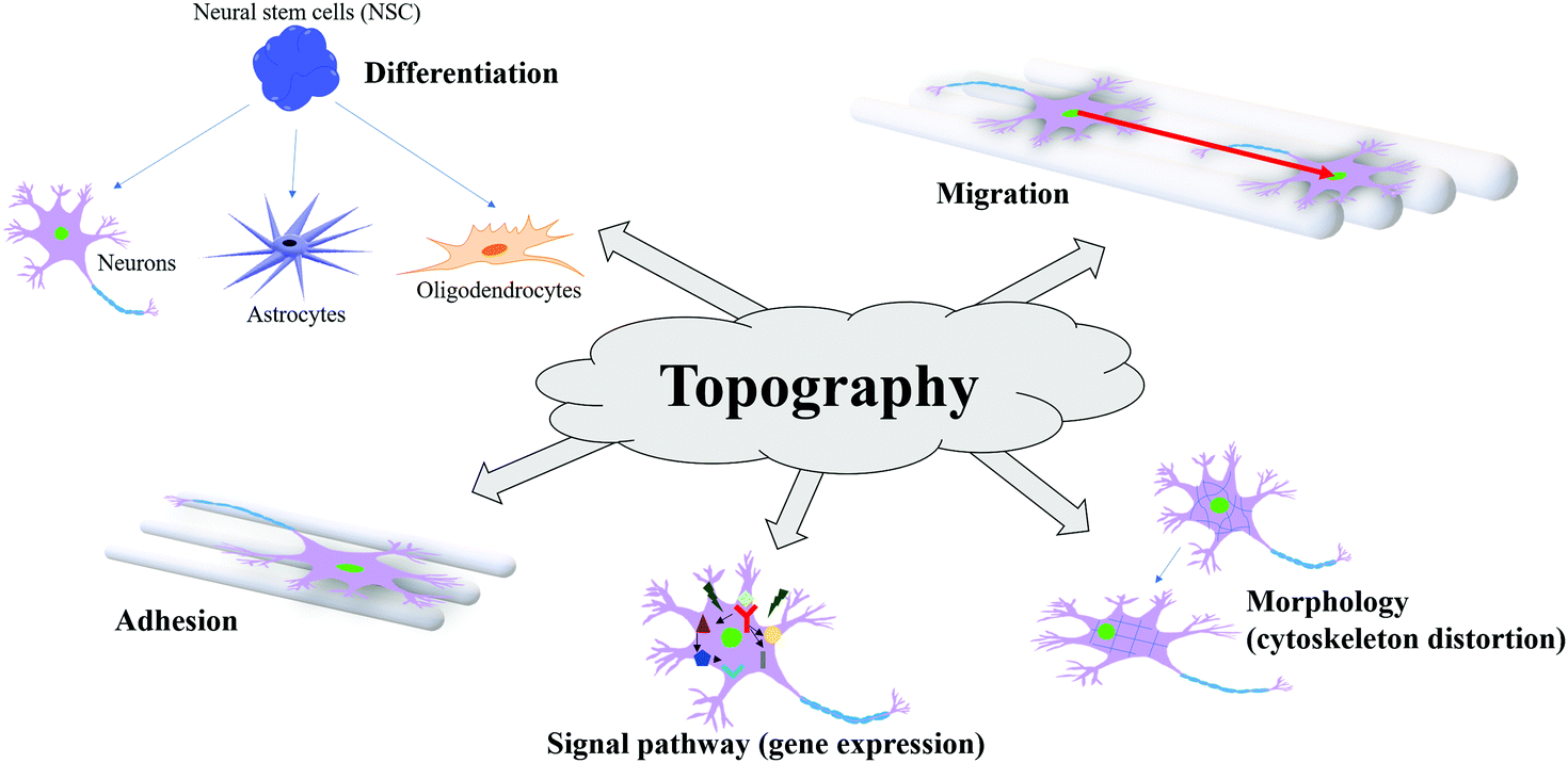

Traditional TE scaffold design strategies employ a ‘top-down’ approach to generate three-dimensional pore structures within biocompatible and biodegradable materials. There are different techniques that can conceivably be used to process biomaterials for porous scaffold development. By considering the wide range of biomaterials and processing techniques for scaffold design, this section will discuss the most relevant aspects related to the processing-pore structure relationships of the scaffolds prepared by some of the most used techniques. Furthermore, recent development in nano-technology have demonstrated that the presence of controlled nano-structures on the surface of biomaterials and scaffolds is essential to promote and guide cellular processes involved in new tissue regeneration, such as cell adhesion, migration and differentiation ( Lim and Donahue, 2007 Ma, 2008 Wang et al., 2011).Ī number of fabrication technologies have been applied to process biocompatible and biodegradable materials into three-dimensional porous scaffolds with controlled nano- to micro-structures, and to ultimately tailor the final biological response. Consequently, to engineer functional tissues and organs successfully, the scaffolds have to be designed with a micro-architecture able to facilitate cell distribution and guide tissue regeneration in three dimensions ( Lutolf and Hubbell, 2005 Guarino et al., 2008).

In natural tissues, cells and ECM are organized into three-dimensional structures from the sub-cellular to the tissue level. Furthermore, design and manufacturing techniques must be used to translate scaffold-based TE from concept to, ultimately, potential clinical applications. Scaffold design and manufacturing are key steps in defining scaffolds with prescribed mechanical, mass-transport and surface characteristics that can be used to test initial hypotheses regarding in vitro cells culture and in vivo tissue regeneration models. Nettis, in Biomedical Foams for Tissue Engineering Applications, 2014 3.2 Micro-structure of biomedical foams and processing techniques Clinical outcomes and complications of a collagen meniscus implant: A systematic review. Grassi A., Zaffagnini S., Muccioli G.M.M., Benzi A., Marcacci M. Actifit (R) polyurethane meniscal scaffold: MRI and functional outcomes after a minimum follow-up of 5 years. Leroy A., Beaufils P., Faivre B., Steltzlen C., Boisrenoult P., Pujol N. Survivorship of Meniscal Allograft Transplantation in an Athletic Patient Population. Waterman B.R., Rensing N., Cameron K.L., Owens B.D., Pallis M. Engineered Healing of Avascular Meniscus Tears by Stem Cell Recruitment.

Tarafder S., Gulko J., Sim K.H., Yang J., Cook J.L., Lee C.H. Influence of Donor Age on the Biomechanical and Biochemical Properties of Human Meniscal Allografts. The 3D-printed hybrid-scaffold may provide a promising approach that can be applied to those who received total meniscal resection, using patient-specific design and autogenous cell source.ģD printing hybrid-scaffold meniscectomy polycaprolactone tissue regeneration.īursac P., York A., Kuznia P., Brown L.M., Arnoczky S.P. Additionally, the hybrid-scaffold did not induce osteoarthritis on the femoral condyle surface. Histological analysis showed evidence of extensive neo-meniscus cell ingrowth. Hybrid-scaffolding resulted in superior shape matching as compared to original meniscus tissue. Histological and immunohistochemical analysis results showed obvious ingrowth of the fibroblast-like cells and chondrocyte cells as well as mature lacunae that were embedded in the extracellular matrix. At postoperative 12-week, hybrid-scaffold achieved neo-meniscus tissue formation, and its shape was maintained without rupture or break away from the knee joint. Based on this hybrid-scaffold with an autograft cell source (fibrochondrocyte), we assessed mechanical and histological results using the rabbit total meniscectomy model. Then, we completed the hybrid-scaffold by combining the 3D-printed framework and mixture micro-size composite which consists of polycaprolactone and sodium chloride to create a cell-friendly microenvironment. We manufactured a polycaprolactone-based patient-specific designed framework via a Computed Tomography scan images and 3D-printing technique. Although there are commercialized off-the-shelf alternatives to achieve total meniscus regeneration, each has its own shortcomings such as individualized size matching issues and inappropriate mechanical properties. The meniscus has poor intrinsic regenerative capability, and its injury inevitably leads to articular cartilage degeneration.


 0 kommentar(er)
